Muscle Attachments
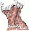
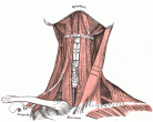
Muscle Images
| Region | Muscle | Origin | Insertion | Action |
|---|---|---|---|---|
| Scalp / Eyelid | Occipitalis | Superior nuchal line of the occipital bone, mastoid part of the temporal bone | Galea aponeurotica | Wrinkles eyebrow |
| Scalp / Eyelid | Frontalis | Galea aponeurotica | Mastoid process | Wrinkles eyebrow |
| Scalp / Eyelid | Orbicularis Oculi | Frontal bone; medial palpebral ligament; lacrimal bone | Lateral palpebral raphe | Closes eyelids |
| Scalp / Eyelid | Corrugator Supercilii | Superciliary arches | Forehead skin, near eyebrow | Wrinkles forehead |
| Scalp / Eyelid | Depressor Supercilii | Medial orbital rim | Medial aspect of bony orbit | Depression of eyebrow |
| Extraocular | Levator Palpebrae Superioris | Sphenoid bone | Tarsal plate, upper eyelid | Retracts / elevates eyelid |
| Extraocular | Superior Tarsal | Underside of levator palpebrae superioris | Superior tarsal plate of the eyelid | Raise the upper eyelid |
| Extraocular | Superior Rectus | Annulus of Zinn at the orbital apex | 7.5 mm superior to the corneal limbus | Elevates, adducts, and rotates medially the eye |
| Extraocular | Inferior Rectus | Annulus of Zinn at the orbital apex | 6.5 mm inferior to the corneal limbus | Depression and adduction |
| Extraocular | Medial Rectus | Annulus of Zinn at the orbital apex | 5.5 mm medial to the corneal limbus | Adducts the eyeball |
| Extraocular | Lateral Rectus | Annulus of Zinn at the orbital apex | 7 mm temporal to the corneal limbus | Abducts the eyeball |
| Extraocular | Superior Oblique | Annulus of Zinn at the orbital apex, medial to optic canal | Outer posterior quadrant of the eyeball | Primary: intorsion. Secondary:abduct (laterally rotate) and depress the eyeball |
| Extraocular | Inferior Oblique | Orbital surface of the maxilla, lateral to the lacrimal groove | Laterally onto the eyeball, deep to the lateral rectus, by a short flat tendon | Extorsion, elevation, abduction |
| Intraocular | Ciliary | Accommodation | ||
| Intraocular | Iris Dilator | Pupil dilation | ||
| Intraocular | Iris Sphincter | Constricts pupil | ||
| Ear | Auriculares | Galeal aponeurosis | Front of the helix, cranial surface of the pinna | (Wiggle ears) |
| Ear | Temporoparietalis | Auriculares muscles | Galea aponeurotica | (Wiggle ears) |
| Ear | Stapedius | Neck of stapes | Control the amplitude of sound waves to the inner ear | |
| Ear | Tensor tympani | Eustachian tube | Handle of the malleus | Tensing the tympanic membrane |
| Nose | Procerus | From fascia over the lower of the nasal bone | Skin of the lower part of the forehead between the eyebrows | Draws down the medial angle of the eyebrow giving expressions of frowning |
| Nose | Nasalis | Maxilla | Nasal bone | Compresses bridge, depresses tip of nose, elevates corners of nostrils |
| Nose | Dilatator Naris | Margin of the nasal notch of the maxilla, greater and lesser alar cartilages | Skin near the margin of the nostril | Dilation of nostrils |
| Nose | Depressor Septi Nasi | Incisive fossa of the maxilla | Nasal septum and back part of the alar part of nasalis muscle | Depression of nasal septum |
| Nose | Levator Labii Superioris Alaeque Nasi | Maxilla | Nostril and upper lip | Dilates the nostril; elevates the upper lip and wing of the nose |
| Mouth | Levator anguli oris | Maxilla | Modiolus of mouth | Smile (elevates angle of mouth) |
| Mouth | Depressor anguli oris | Tubercle of mandible | Modiolus of mouth | Depresses angle of mouth |
| Mouth | Levator labii superioris | Medial infra-orbital margin | Skin and muscle of the upper lip (labii superioris) | Elevates the upper lip |
| Mouth | Depressor labii inferioris | Oblique line of the mandible, between the symphysis and the mental foramen | Integument of the lower lip, Orbicularis oris fibers, its fellow of the opposite side | Depress the lower lip |
| Mouth | Zygomaticus major | Anterior of zygomatic | Modiolus of mouth | Draws angle of mouth upward and laterally |
| Mouth | Zygomaticus minor | Zygomatic bone | Skin of the upper lip | Elevates upper lip |
| Mouth | Mentalis | Anterior mandible | Chin | Elevates and wrinkles skin of chin, protrudes lower lip |
| Mouth | Buccinator | Alveolar processes of the maxillary bone and mandible, pterygomandibular raphe | In the fibres of the orbicularis oris | Compress the cheeks against the teeth (blowing), mastication. |
| Mouth | Orbicularis oris | Maxilla and mandible | Skin around the lips | Pucker the lips |
| Mouth | Risorius | Parotid fascia | Modiolus | Draw back angle of mouth |
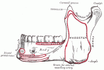
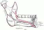
| Mouth | Masseter | Zygomatic arch and maxilla | Coronoid process and ramus of mandible | Elevation (as in closing of the mouth) and retraction of mandible |
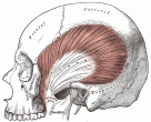
| Mouth | Temporalis | Temporal lines on the parietal bone of the skull | Coronoid process of the mandible | Elevation and retraction of mandible |
| Mouth | Lateral Pterygoid | Great wing of sphenoid and pterygoid plate | Condyle of mandible | Depresses mandible |
| Mouth | Medial Pterygoid | Deep head: medial side of lateral pterygoid plate behind the upper teeth; superficial head: pyramidal process of palatine bone and maxillary tuberosity | Medial angle of the mandible | Elevates mandible, closes jaw, helps lateral pterygoids in moving the jaw from side to side |
| Tongue - Extrinsic | Genioglossus | Superior part of mental spine of mandible (symphysis menti) | Dorsum of tongue and body of hyoid | Complex - Inferior fibers protrude the tongue, middle fibers depress the tongue, and its superior fibers draw the tip back and down |
| Tongue - Extrinsic | Hyoglossus | Hyoid | Side of the tongue | Depresses tongue |
| Tongue - Extrinsic | Chondroglossus | Lesser cornu and body of the hyoid bone | Intrinsic muscular fibers of the tongue | Depresses tongue (some consider this muscle to be part of hyoglossus) |
| Tongue - Extrinsic | Styloglossus | Styloid process of temporal bone | Tongue | Elevates and retracts tongue |
| Tongue - Intrinsic | Superior longitudinal | Close to the epiglottis, from the median fibrous septum | Edges of the tongue | Retracts the tongue with the inferior longitudinal muscle, making the tongue short and thick |
| Tongue - Intrinsic | Transversus | Median fibrous septum | Sides of the tongue | Makes the tongue narrow and elongated |
| Tongue - Intrinsic | Inferior longitudinal | Root of the tongue | Apex of the tongue | |
| Soft Palate | Levator veli palatini | Temporal bone, Eustachian tube | Palatine aponeurosis | Elevates soft palate |
| Soft Palate | Tensor veli palatini | Medial pterygoid plate of the sphenoid bone | Palatine aponeurosis | Tension of the soft palate |
| Soft Palate | Musculus uvulae | Hard palate | Palatine aponeurosis | |
| Soft Palate | Palatoglossus | Palatine aponeurosis | Tongue | Raising the back part of the tongue |
| Soft Palate | Palatopharyngeus | Palatine aponeurosis and hard palate | Upper border of thyroid cartilage (blends with constrictor fibers) | Pulls pharynx and larynx |
| Pharynx | Pharyngeal Constrictor Inferior | Cricoid and thyroid cartilage | Pharyngeal raphe | Swallowing |
| Pharynx | Pharyngeal Constrictor Middle | Hyoid bone | Pharyngeal raphe | Swallowing |
| Pharynx | Pharyngeal Constrictor Superior | Medial pterygoid plate, pterygomandibular raphé, alveolar process | Pharyngeal raphe, pharyngeal tubercle | Swallowing |
| Pharynx | Stylopharyngeus | Styloid process (temporal) | Thyroid cartilage (pharynx) | Elevate the larynx, elevate the pharynx, swallowing |
| Pharynx | Salpingopharyngeus | Cartilage of the Eustachian tube | Posterior fasciculus of the pharyngopalatinus muscle | Raise the nasopharynx |
| Larynx | Cricothyroid | Anterior and lateral cricoid cartilage | Inferior cornu and lamina of the thyroid cartilage | Tension and elongation of the vocal folds (has minor adductory effect) |
| Larynx | Cricoarytenoid Posterior | Posterior part of the cricoid | Muscular process of the arytenoid cartilage | Abducts and laterally rotates the cartilage, pulling the vocal ligaments away from the midline and forward and so opening the rima glottidis |
| Larynx | Cricoarytenoid Lateral | Lateral part of the arch of the cricoid | Muscular process of the arytenoid cartilage | Adduct and medially rotate the cartilage, pulling the vocal ligaments towards the midline and backwards and so closing off the rima glottidis |
| Larynx | Arytenoid | Arytenoid cartilage on one side | Arytenoid cartilage on opposite side | Approximate the arytenoid cartilages (close rima glottidis) |
| Larynx | Thyroarytenoid | Inner surface of the thyroid cartilage (anterior aspect) | Anterior surface of arytenoid cartilage | Thickens the vocal folds and decreases length; also helps helps to adduct the vocal folds during speech |

