
Shoulder Fractures – Imaging and Management
Accordion Sample Description
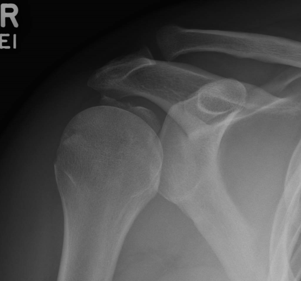

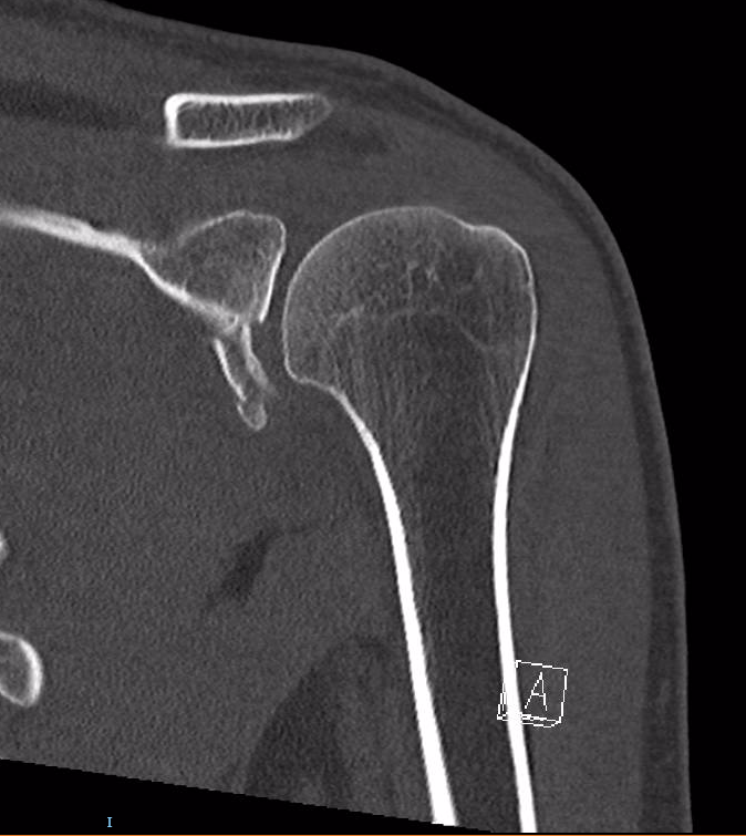
Glenoid fractures are commonly associated with glenohumeral dislocations. These may appear subtle on x-rays and may need further imaging such as CT scan to quantify.
These images show a fracture to the inferior glenoid sustained after a shoulder dislocation, termed a "Bankart" fracture. Depending on the displacement of the fracture, management might range from a period of immobilisation to surgery. Orthopaedic opinion is usually warranted.


Accordion Sample Description
Dislocations and Subluxations About The Shoulder Girdle – Imaging and Management
Accordion Sample Description
- WIth a posterior glenohumeral dislocation, on the AP x-ray there is the appearance of the "Globe" sign, where because the humerus is rotated internally, it appears like a lightglobe.
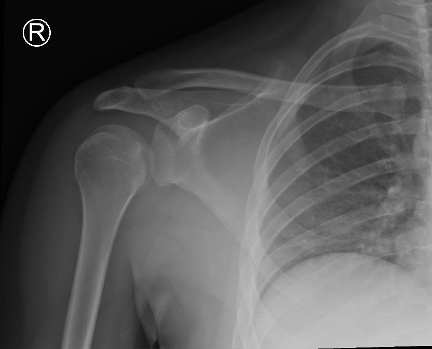
- On the lateral, the centre of the humerus is located out of the Y-shaped "propeller" formed by the coracoid process, acromion and blade of the scapula

Soft Tissue Injuries About The Shoulder – Imaging and Management
Accordion Sample Description
Accordion Sample Description